

Please wait...
About This Project
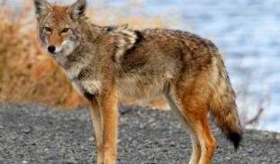
Rocky Mountain spotted fever (RMSF) is a tick-borne bacterial infection that can cause permanent disability or death in humans, with a case fatality rate up to 28% untreated. In 2003, RMSF moved into Arizona, where it is vectored by the brown dog tick (Rhipicephalus sanguineus).
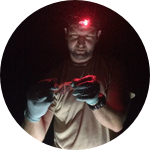
We would like to determine whether coyotes develop antibodies for RMSF - indicating that they are affected by the disease - and investigate whether they facilitate the dispersal of RMSF-infected ticks.
More Lab Notes From This Project

Browse Other Projects on Experiment
Related Projects
Do Australian bats have what it takes to survive the deadly White Nose Syndrome?
Australian bats are at risk from the deadly fungal disease White Nose Syndrome (WNS), which is expected...
Are climate change and air pollution triggering cardiovascular disease?
Ongoing global climate change and air pollution emissions pose a major threat to cardiovascular health...
Can blood lactate levels help guide treatment for birds suffering from monofilament line entanglement injuries?
Fishing line entanglement injuries are a common problem affecting over 200 different wildlife species globally...