


Please wait...
About This Project
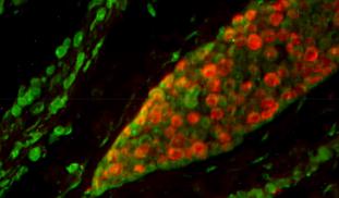
Cancer treatment is a challenging, complex and exhausting process for the patient, their family and medical team. Chemotherapy kills both cancer and normal cells, and may not prevent remaining cancer cells from spreading. Our research team believes that by turning up a gene HEXIM1, it has a way to stop most cancers from spreading. This could make cancer treatment more palatable and effective and ultimately make cancer a more survivable disease.

Browse Other Projects on Experiment
Related Projects
Molecular target-based focused screen for Chagas' disease therapeutic drug discovery
The etiological agent for Chagas' disease is the protozoan parasite Trypanosoma cruzi, which affects 6...
Target-based drug discovery for coronavirus disease 2019
Therapeutics in any modality to combat Severe Acute Respiratory Syndrome Coronavirus 2 (SARS-CoV-2) infections...
Early-Stage Drug Discovery for Chagas' Disease
An estimated 6 – 7 million people in Latin America and 300,000 U.S. citizens are infected with the protozoan...