
Please wait...
About This Project
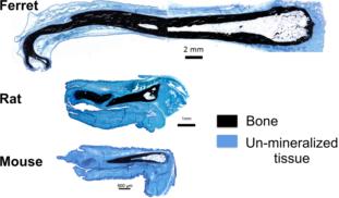
Most mammals have penis bones, also called bacula. Our recent review indicated that penis bones evolved 9 independent times. Based on this study, the origin of a rodent penis bone is separate from the origin in carnivores. This got us thinking, do these independent events utilize the same set of genes to develop penis bones? In this project we will investigate the expression of a critical bone development gene across species that evolved bacula independently.
