


Please wait...
About This Project
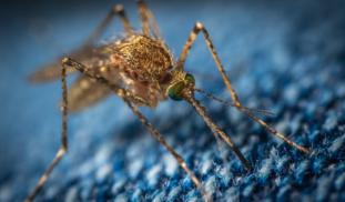
As of mid-2019, 87 countries have had or still have Zika cases, underlining the importance of this infectious disease. Zika virus can infect the uterus and later infect the infant during pregnancy, causing neurodevelopmental defects at birth. This study will model Zika infection in the lab using uterine mini-organs. We hypothesize that our specific antibodies can neutralize Zika virus in the uterus and thus prevent later transmission from pregnant mother to unborn child.

Browse Other Projects on Experiment
Related Projects
Shutting down cancer’s recycling system with exosome-based therapy
Pancreatic cancer is one of the deadliest cancers because its cells survive by recycling their own components...
Developing a novel oxysterol antibiotic to combat drug-resistant tuberculosis
Drug-resistant tuberculosis (TB) is a consistently growing threat to global health. We have developed Oxy291...
Tote-Size portable incubator for rapid field work
Waiting for lab results is slowing science down! We are designing a fully open source portable incubator...