455
0
0
Like?
Please wait...
About This Project
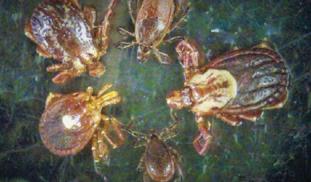
To “show one's metal” is to reveal one’s strength of character. Ticks may have tiny character, but I propose they do have metal! As blood sucking parasites, ticks must penetrate thick animal skin to obtain food. I propose that ticks enrich their appendages with metals to strengthen them for penetration. I intend to use electron microscopy to quantify the chemical elements in the appendages of deer, dog, and lone star ticks to observe their composition and determine if ticks employ metals.
More Lab Notes From This Project

Browse Other Projects on Experiment
Related Projects
Out for blood: Hemoparasites in white-tailed deer from the Shenandoah Valley in Northern Virginia
Our research question centers about the prevalence and diversity of hemoparasites that infect ungulate poplulations...
Using eDNA to examine protected California species in streams at Hastings Reserve
Hastings Reserve is home to three streams that provide critical habitat for sensitive native species. Through...
How do polar bears stay healthy on the world's worst diet?
Polar bears survive almost entirely on seal fat. Yet unlike humans who eat high-fat diets, polar bears never...

