26
0
0
Like?
Please wait...
About This Project
University of Massachusetts Lowell
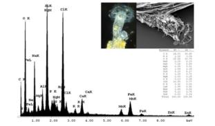
Energy dispersive x-ray spectroscopy (EDS) is a method for analyzing the elemental (chemical) composition of synthetic and natural structures, and has been useful in identifying trace metals in forensics and ecotoxicology. To date, it has never been applied to detect trace elements in invertebrate secretions. Here, I describe a study that uses EDS to characterize the protective secretions of sessile rotifers and to determine if metal pollutants are incorporated into their secretions.

Browse Other Projects on Experiment
Related Projects
Out for blood: Hemoparasites in white-tailed deer from the Shenandoah Valley in Northern Virginia
Our research question centers about the prevalence and diversity of hemoparasites that infect ungulate poplulations...
Using eDNA to examine protected California species in streams at Hastings Reserve
Hastings Reserve is home to three streams that provide critical habitat for sensitive native species. Through...
How do polar bears stay healthy on the world's worst diet?
Polar bears survive almost entirely on seal fat. Yet unlike humans who eat high-fat diets, polar bears never...

