About This Project
Energy dispersive x-ray spectroscopy (EDS) is a method for analyzing the elemental (chemical) composition of synthetic and natural structures, and has been useful in identifying trace metals in forensics and ecotoxicology. To date, it has never been applied to detect trace elements in invertebrate secretions. Here, I describe a study that uses EDS to characterize the protective secretions of sessile rotifers and to determine if metal pollutants are incorporated into their secretions.
Ask the Scientists
Join The DiscussionWhat is the context of this research?
Rotifers have a long history of use in ecotoxicology, but those studies generally focus on planktonic species. Importantly, not all toxic compounds remain in the plankton and many settle to the benthos and may accumulate in sediments and on submerged plants. A wide variety of invertebrates, including sessile rotifers, live on these plants and have been shown to collect trace metals in the defensive tubes they secrete around their bodies. Whether these metals are passively or actively incorporated into the secretions remains unknown. Here, I propose to use EDS to characterize the elemental makeup of the protective secretions of sessile rotifers from a variety of polluted and non-polluted ponds in New England, with a focus on the detection of heavy metals such as mercury and arsenic.
What is the significance of this project?
Sessile rotifers live on submerged aquatic plants in freshwater ponds and lakes. As adults, these rotifers are strictly sessile and cannot move once settled, meaning that any pollution in their environment cannot be avoided. Most species secrete protective tubes around their bodies to ward off predators and my preliminary studies have shown these tubes to collect trace metals (As, Hg) from surrounding water. The proposed research is an extension of these studies. First, I will use EDS to determine the elemental composition of rotifer tubes from unpolluted and polluted ponds in New England, the latter of which have a history of residential and agricultural pollution. Second, I will set up experiments to determine if trace metals are actively or passively incorporated into their tubes.
What are the goals of the project?
I will collect sessile rotifers from lakes and ponds throughout New England for EDS analysis. Species from the field will be used for two purposes. First, females will be cultured to collect larvae that can be transferred to water of predetermined chemistry (i.e., with and without pollutants). These larvae will be placed in synthetic pond water and fed cultured algae. Water chemistry will be altered by adding trace amounts of heavy metals (arsenic, cadmium, mercury, nickel) that are representative of some contaminants in New England water bodies. These animals, as well as field-collected specimens, will then be processed for EDS to determine the ratios of heavy metals to other elements (e.g., carbon, nitrogen, oxygen) that make up their defensive secretions.
Budget
We are requesting funds for the preparation and analysis of rotifer samples on a JEOL JSM 7401F Field Emission Scanning Electron Microscope equipped for X-ray microanalysis. Funds are requested for the following:
1. SEM stub mounts and carbon tap: $50
2. Use of the Au sputter coater: $20/hr for four hours = $80
3. Use of the JEOL JSM-7401F at the Materials Characterization Laboratory at the University of Massachusetts Lowell. Self use at $74/hr x 20 hours and assisted use at $270/hr for 2 hours to learn the EDAX Genesis XM2 Imaging System for elemental microanalysis. Total: $2,020.
 Project Timeline
May 28, 2017
Collect and culture sessile rotifers from Mascuppic Lake Dam, Tyngsboro, MA.
Jun 11, 2017
Collect and culture sessile rotifers from Ayers Pond, Hudson, NH.
Jun 25, 2017
Collect and culture sessile rotifers from Attitash Lake, Amesbury, MA.
Jul 02, 2017
Begin prepping and processing samples for EDS/FE-SEM analysis.
Jun 04, 2018
Present findings at the XV International Rotifer Symposium, University of Texas, El Paso.
Meet the Team
Affiliates
Adele Hochberg
I am an invertebrate zoologist who traditionally works on obscure, under-studied aquatic invertebrates, such as rotifers. I received my B.S. and M.S. in Biological Sciences from the University of Massachusetts Lowell, where I focused on the functional morphology of sessile, gnesiotrochan rotifers and began preliminary investigations of using non-model rotifer species (e.g., sessile, gnesiotrochan rotifers) as bio-indicators of pollution (e.g., metals, etc.) in local aquatic systems. I am currently in my first year as a PhD student at the University of Massachusetts Boston.
Additional Information
EDS is a technique used in conjunction with the field emission scanning electron
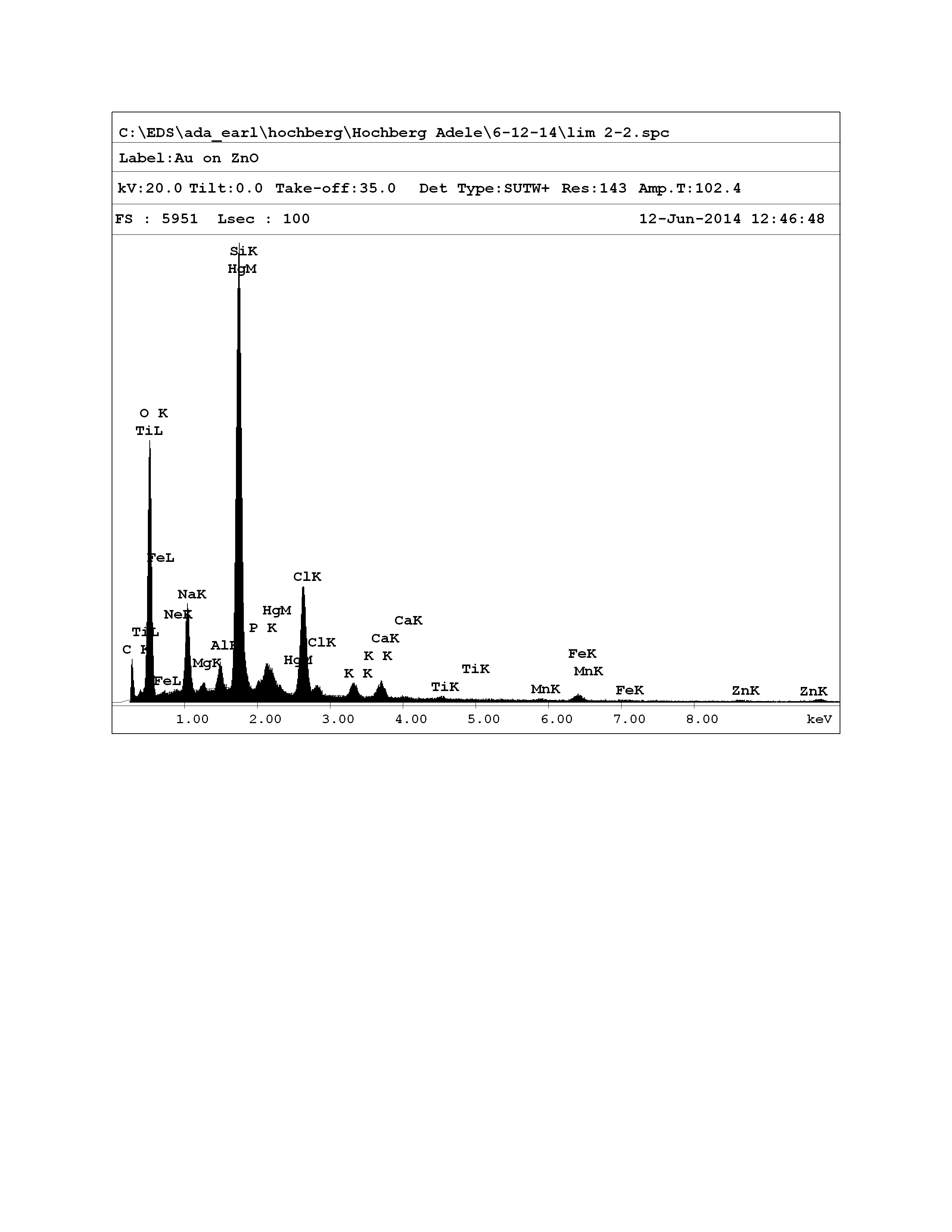
microscope (FE-SEM). When the electron beam from the FE-SEM hits the sample, it ejects an e- from an inner shell (e.g., K shell, see below) that is replaced with an e- from a higher-energy outer shell. The difference in energy levels between these electrons is given off as a characteristic x-ray. The x-rays are then displayed graphically as an EDS spectrum (see figure, below). Each element and its corresponding shells have their own diagnostic x-ray spectral peaks that are easily visualized. Taking the integral of the peaks in the spectrum gives the relative abundance of each element present in the scanned area, providing both the atomic and weight percentages for each element. However, the voltage of the electron beam limits how far the beam can penetrate into the specimen. For example, a 20kV beam penetrates 4.10 μm of pure carbon but only 0.5 μm of pure gold. A normally coated specimen for FE-SEM is covered by only several Å of gold, meaning that an electron beam will penetrate both the gold coating and approximately 4 μm of specimen.
Project Backers
- 0Backers
- 0%Funded
- $0Total Donations
- $0Average Donation